
Lung Cancer and Bronchoscopy
Lung Cancer
UCSF provides state-of-the-art care to patients with lung cancer and cancer that has metastasized to the chest. Our pulmonologists evaluate patients who have lung nodules, lung masses or symptoms suggestive of lung cancer. We can help to determine whether a patient has lung cancer, to help to manage the symptoms associated with lung cancer, and to provide ongoing care after treatment for lung cancer.
Lung Cancer Screening
Screening with annual low dose CT scan of the chest in high risk individuals in one large randomized controlled trial (National Lung Screening Trial) was associated with a 20% decrease in lung cancer mortality and a 6.7% decrease in all-cause mortality. UCSF Medical Center offers a Lung Cancer Screening Program for qualified candidates. Please see the Lung Cancer Screening Program website for more information.
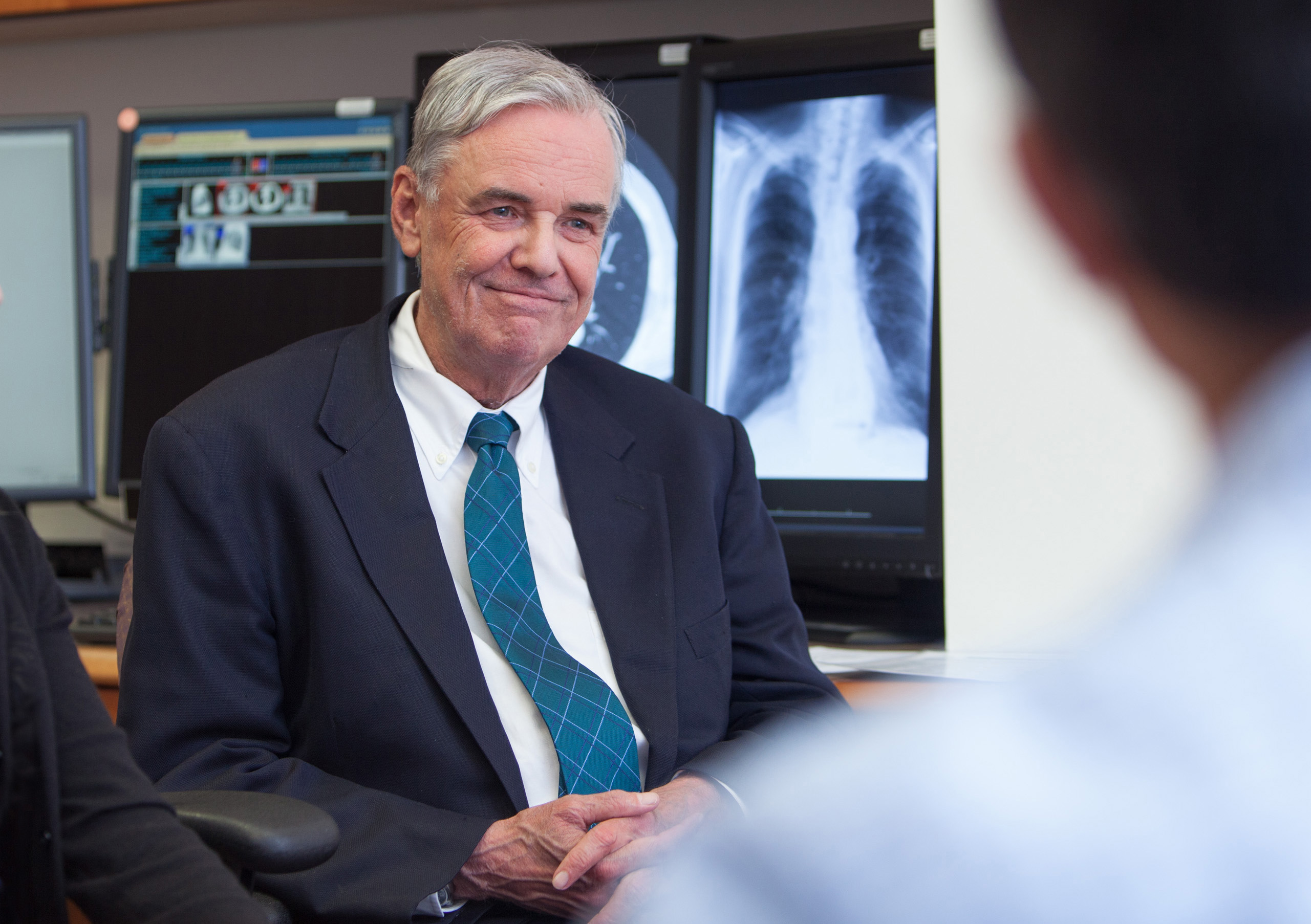
Meet the Team
Patients receive state-of-the-art care from a multidisciplinary team of dedicated to treating lung cancer, mesothelioma, esophageal cancer, metastasiic disease to the lungs as well as less common malignancies including sarcomas, and tumors of the thymus, mediastinum, bronchus and chest wall. In addition, clinical investigators in the program conduct clinical trials for the most promising new therapies. Learn more about the team here.
Our Lung Cancer Pulmonologist Experts include:
- Lorriana Leard, MD –Specializing in Pulmonary Nodules, Lung Cancer Screening, and Advanced Bronchoscopic Procedures
- James Brown, MD - Specializing in VA lung nodule program
- Neil Trivedi, MD - Specializing in Advance Bronchoscopic Procedures
- Eric Seeley, MD - Specializing in Interventional Pulmonology
- Yaron Gesthalter, MD - Specializing in Interventional Pulmonology
Bronchoscopic Procedures
Bronchoscopy is a procedure that is used to view the trachea and bronchial tubes and is used to diagnose lung disease and infection. It may also be used during the treatment of some lung conditions.
Advanced Bronchoscopic Procedures
UCSF Pulmonologists perform some of the most advanced bronchoscopic procedures that can be used to diagnose lung cancer, to determine the clinical stage, or to assist with treatment. These procedures include:
- Endobronchial Ultrasound (EBUS).
- Electromagnetic Navigation Guided Bronchoscopy.
- Stent Placement.
- Fiducial Marker placement to guide Stereotactic body radiation therapy (SBRT).
- Rigid Bronchoscopy: used for foreign body removal and complex airway procedures.
- Endobronchial valve placement: for lung volume reduction in COPD/Emphysema and bronchopleural fistulas
UCSF pulmonologists work very closely with a team of Thoracic Surgeons, Thoracic Oncologists, and Thoracic Radiation Oncologists to offer the most advanced treatment possible.
How the Test is Performed
The procedure is performed using a flexible bronchoscope. A bronchoscope is thin device, usually less than 1/2 inch wide and 2 feet long, which has a small camera on the end to allow the operator to see deep into the lungs. The bronchoscope also has a channel that can be used to pass small instruments or fluid into the airway to collect samples.
When the scope is in the area of interest the doctor will take some samples using any of the following techniques.
- Bronchoalveolar Lavage: During this procedure the physician passes saline solution through the scope and into the airway and out to the lung. The fluid is then suctioned back through the scope into a container where it can be sent to the lab for diagnostic testing.
- Transbronchial Biopsies: During this procedure a small forcep is passed through the scope into the lung and small samples of lung tissue are removed. This tissue can then be sent to the lab for diagnostic testing.
- Transbronchial Needle Aspiration: During this procedure a small needle is passed through the bronchoscope and through the bronchial wall to sample lymph node or lung tissue.
- Brush Biopsy: During this procedure a small brush is passed through the scope and into the airway where the brush can collect cells from an area of the airway wall. The brush sample can then be sent to the lab for diagnostic testing.
There are other devices that may be used during bronchoscopy. These include:
- Endobronchial Ultrasound: Using this tool, the physician can identify lymph nodes or abnormal areas of the lung using a small ultrasound probe that is attached to the end of a special bronchoscope
- Electromagnetic Navigational Bronchoscopy: Using this tool, the physician will use special equipment that allows 3D reconstruction of the bronchial tubes and development of a pathway that helps guide the physician to an area of interest in the lungs to allow for sampling.
How to Prepare for the Test
Do not eat or drink anything at least 6 hours prior to the procedure. You should not take blood thinning medication such as Coumadin or Heparin shots prior to the procedure.
You will need to arrange to have someone pick you up from the bronchoscopy recovery area and drive you home.
What to Expect Following the Test
Following the procedure, you will not have a normal gag reflex for 1-2 hours after the test. Therefore you will not be able to eat or drink during this time.
When the anesthetic wears off, your throat may be scratchy for a few days.
You will likely have an increased cough for a few days following the procedure.
You may have a low grade fever during the 24 hours following the procedure.
Why the Test is Performed
You may have a bronchoscopy to help your doctor diagnose lung problems. Your doctor will be able to inspect the bronchial tubes or take a sample of your lung tissue.
Common reasons to perform a bronchoscopy for diagnosis are:
- Lung growth, lymph node, atelectasis, or other changes seen on an x-ray or other imaging test
- Suspected interstitial lung disease
- Coughing up blood (hemoptysis)
- Possible foreign object in the airway
- Cough that has lasted more than 3 months without any other explanation
- Infections in the lungs and bronchi that cannot be diagnosed any other way or need a certain type of diagnosis
- Inhaled toxic gas or chemical
- To diagnose a lung rejection after a lung transplant
Risks
The physician will go through in the detail the risks associated with bronchoscopy. In general, this procedure is very safe and associated with complications very rarely.
The main risks from bronchoscopy are:
- Bleeding from biopsy sites
- Infection
- Pneumothorax is when a small hole is created in the lining of the lung